
Most FRCEM candidates are confident with the obvious ECG presentations — the anterior STEMI with textbook ST elevation, the VF trace before defibrillation, the classic hyperkalaemic sine wave. Those aren't what trips people up in the exam.
What trips people up are the patterns that look deceptively quiet. The ECG that doesn't scream emergency. The trace where the T waves are just a little different, or there's a subtle right precordial change that doesn't meet classic STEMI criteria — but the patient is about to arrest.
These are the silent killers. FRCEM tests them deliberately, because recognising them is exactly what separates a safe emergency physician from a dangerous one. Miss these patterns in real life and patients die. Miss them in the SBA and you lose marks on questions you should have scored.
This guide covers five high-yield, high-stakes ECG patterns that FRCEM tests consistently — with exactly what to look for, why they matter, and what you do when you see them.
Why ECG Pattern Recognition Is Tested So Heavily in FRCEM
ECG interpretation threads through SLO1, SLO3, and SLO4. It's not a standalone topic — it's embedded in ACS questions, resuscitation scenarios, toxicology cases, and metabolic emergency presentations. Getting fast and accurate at pattern recognition on an ECG trace is one of the highest-leverage skills you can develop for this exam.
The patterns below are specifically chosen because they share two features: they're dangerous if missed, and they're regularly tested precisely because many candidates have a blind spot around them.
If you need to revisit the core STEMI versus NSTEMI framework first, the FRCEM ECG and ACS guide covers territory identification, ST elevation criteria, and standard management timing. This guide builds beyond that foundation into the patterns that sit outside the obvious.
1. Wellens' Syndrome — The Pre-Infarction Time Bomb
What It Is
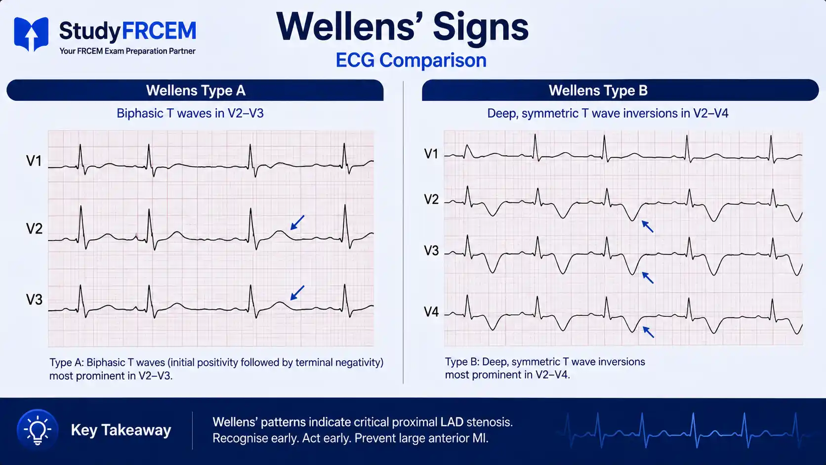
Wellens' syndrome describes a pattern of electrocardiographic changes, particularly deeply inverted or biphasic T waves in leads V2–V3, that is highly specific for critical proximal stenosis of the left anterior descending coronary artery. It signals a pre-infarction state — the LAD is critically narrowed and the patient is heading toward a massive anterior MI, possibly within days.
Typically when patients with Wellens syndrome present to the emergency department they are pain-free, and cardiac enzymes are normal or only slightly elevated. This is the core exam trap: the patient isn't in pain, the troponin is near-normal, and the ECG looks like just "some T wave changes." Nothing about the presentation screams emergency — except the pattern itself.
When Wellens and colleagues first identified the syndrome, they found that 75% of patients with these ECG findings went on to develop acute anterior wall myocardial infarction within weeks when treated with medical management alone — which is why recognition and urgent intervention are non-negotiable.
The Two Patterns
Two distinct ECG patterns define Wellens' syndrome: biphasic T waves in V2–V3 (Type A) or deeply negative T waves in V2–V4 (Type B).
Type A (25% of cases): Biphasic T waves in V2–V3 — less common but more specific. The T wave goes positive then negative (or negative then positive). Easy to dismiss as "non-specific ST-T changes."
Type B (75% of cases): Deep, symmetrical T wave inversions in V2–V4 — the more common pattern. These are deeply inverted, symmetric, and clearly abnormal in the anterior leads.
Both patterns appear during pain-free intervals — which is exactly why they get missed. The patient has settled, the troponin isn't particularly dramatic, and the temptation is to triage them lower.
What FRCEM Tests
A scenario where a patient presented with chest pain hours ago, has now settled with no pain, has a mildly elevated or normal troponin, and the ECG shows deep symmetric T wave inversions in the anterior leads. The question asks: what is the most appropriate next step?
The wrong answers: discharge with outpatient follow-up, exercise stress test, repeat troponin at 6 hours.
The moment Wellens syndrome is suspected, the correct pathway is urgent coronary angiography — not a stress test, not outpatient follow-up.
Stress testing is specifically contraindicated — increasing cardiac demand in a patient with 95% proximal LAD stenosis risks triggering the very anterior MI you're trying to prevent.
Correct answer: Urgent cardiology referral for coronary angiography. Admit, do not discharge, do not stress test.
2. De Winter's T-Wave Pattern — The STEMI That Doesn't Look Like One
What It Is
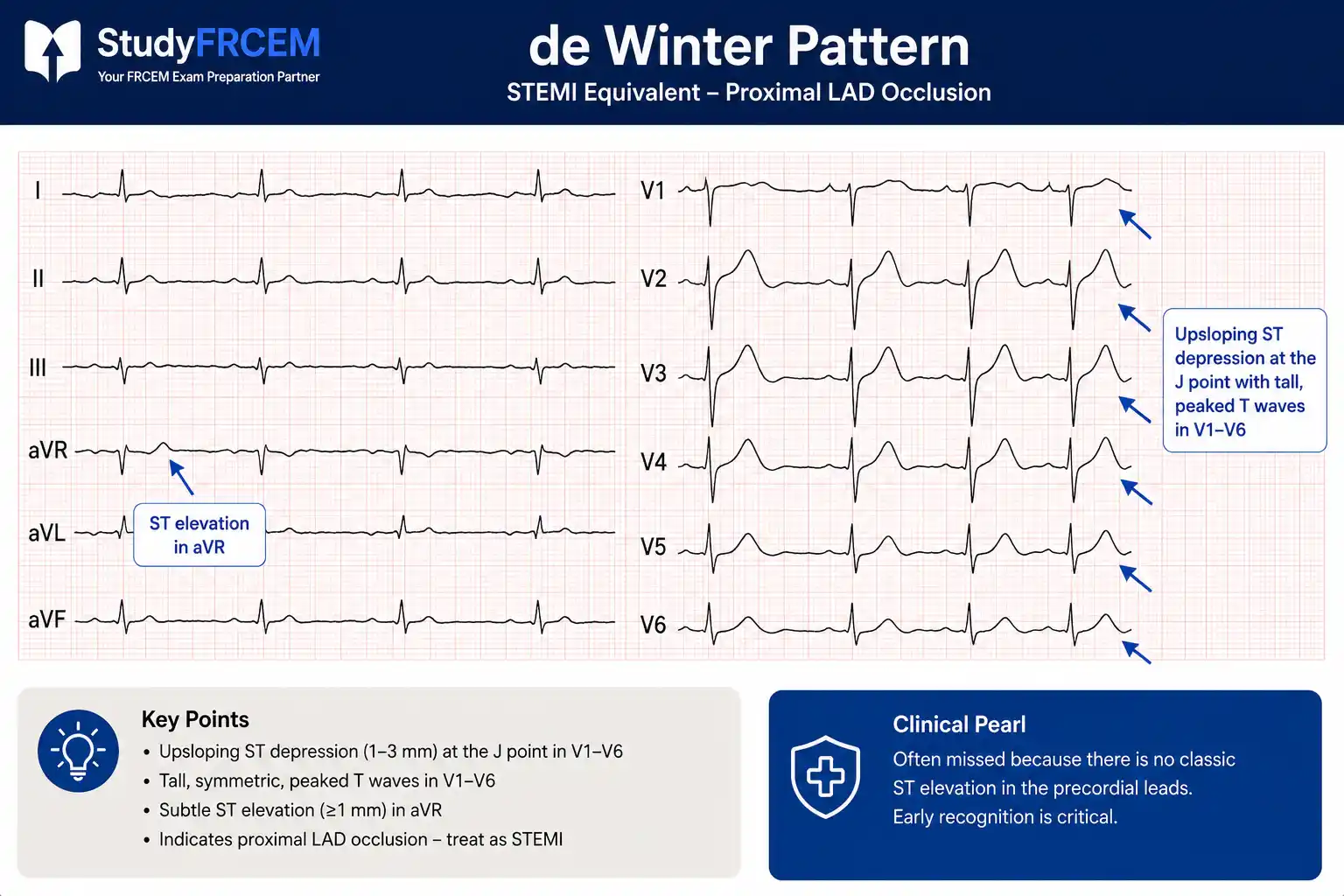
De Winter's T-wave pattern represents acute proximal LAD occlusion — functionally identical to an anterior STEMI in terms of urgency and management — but it lacks the classic ST elevation that would trigger a STEMI alert on most automated ECG systems and in most candidates' minds.
The ability to recognise de Winter's pattern is critical — definitive treatment is urgent cardiac catheterisation with PCI, and failure to identify it leads to the same outcome as a missed anterior STEMI: potentially catastrophic myocardial loss.
What to Look For
The de Winter pattern has three characteristic features:
Upsloping ST depression at the J-point in the precordial leads (V1–V6) — usually 1–3 mm
Tall, peaked, prominent T waves in the same leads — the T waves are large and symmetric
Subtle ST elevation in aVR — often a few millimetres, easy to overlook
There is no ST elevation in the anterior leads. The automated "STEMI detected" prompt won't fire. Machines miss it. Candidates miss it. That's why it's tested.
The underlying pathophysiology: the pattern occurs when anterior ischaemia creates a specific electrophysiological state that manifests as depression and tall T waves rather than elevation — but the occlusion and myocardial threat are identical to anterior STEMI.
What FRCEM Tests
A scenario presenting a patient with acute chest pain and an ECG showing upsloping ST depression with tall T waves in the precordial leads — no ST elevation anywhere. The question asks what this represents and what should happen next.
Answer: De Winter's T-wave pattern — STEMI equivalent. Immediate cath lab activation. Treat identically to anterior STEMI.
This is a STEMI equivalent and is listed as such in the latest ESC ACS guidelines. The management pathway — immediate primary PCI — is identical to a conventional STEMI. Do not manage as NSTEMI.
3. Brugada Pattern — Sudden Cardiac Death in a Seemingly Well Patient
What It Is
First described in 1992 in previously healthy patients who presented with unprovoked syncope and then developed polymorphic ventricular tachycardia — either self-terminating or fatal — Brugada syndrome remains one of the most important causes of sudden cardiac death in structurally normal hearts.
It is an inherited channelopathy — sodium channel dysfunction creating a characteristic right precordial ECG pattern — with no abnormality visible on echocardiography. Patients are typically young to middle-aged adults, often male, and may present with syncope, palpitations, or an apparently incidental ECG finding. Recognising this pattern in the ED is important because several common triggers can unmask it — and those triggers are correctable.
The Pattern: Only Type 1 Is Diagnostic
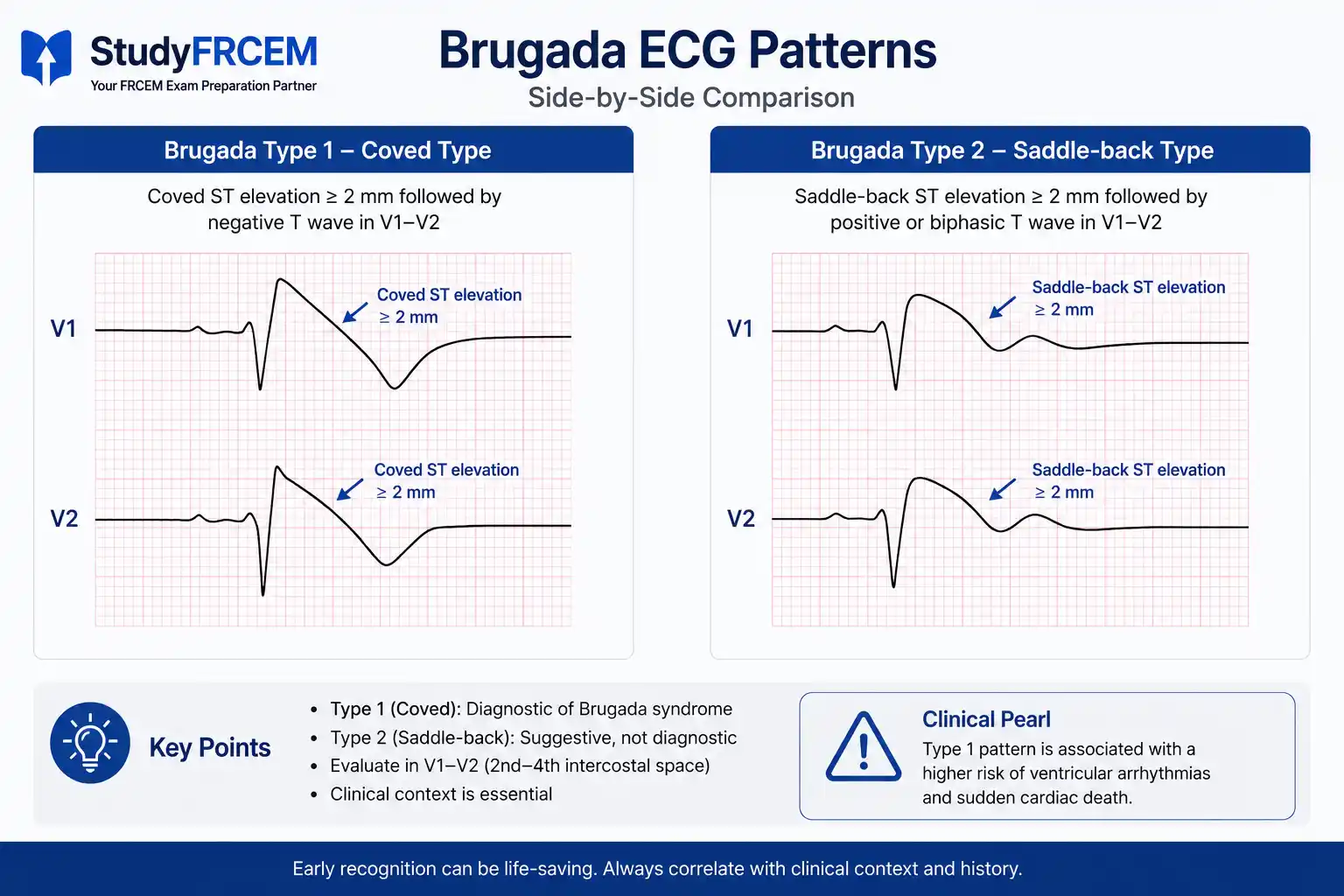
Three ECG pattern types exist, but only Type 1 is considered diagnostic of Brugada syndrome.
Type 1 (diagnostic — coved pattern):
An rS complex followed by at least 2mm of concave ST elevation which descends into an inverted and symmetric T wave in V1–V3.
This "coved" appearance — ST elevation curving downward into T wave inversion — in V1–V3 (or high right precordial leads V1–V2 at the 2nd intercostal space) is the diagnostic pattern.
Type 2 (saddle-back pattern):
An rSr' complex where the r' is at least 2mm, giving a saddle-back ST-segment morphology.
Not diagnostic on its own. Requires provocation testing (sodium channel blocker challenge) to determine if underlying type 1 pattern is present.
For FRCEM: Know that type 1 is the only diagnostic pattern. Type 2 raises suspicion but is not confirmatory.
Triggers That Unmask the Pattern
The Brugada ECG can be dynamic — the pattern comes and goes. Several factors can unmask or worsen it:
Fever — one of the most common ED triggers; a febrile patient with coved right precordial changes may have fever-unmasked Brugada. Treat the fever aggressively with antipyretics and monitor for arrhythmia.
Certain drugs: sodium channel blockers (flecainide, propafenone, tricyclics), some antipsychotics
Electrolyte abnormalities — particularly hypokalaemia and hypercalcaemia
Alcohol excess and large meals
What FRCEM Tests
Two scenario types appear:
Scenario 1: A patient with syncope has an ECG showing coved ST elevation in V1–V2. They're currently well. The question asks about the pattern and management.
Answer: Type 1 Brugada pattern — refer for urgent cardiology review and electrophysiology evaluation. Do not discharge without cardiology involvement. Avoid sodium channel–blocking drugs.
Scenario 2: A febrile patient has an ECG showing coved right precordial changes.
Answer: Fever-induced Brugada unmasking. Treat the fever actively (antipyretics), monitor, and refer to cardiology. Recognise that the ECG may normalise when the fever resolves — but the underlying syndrome may be present.
ED Management of Brugada with VT/VF: Defibrillation for VF as per ALS algorithm. Isoprenaline or quinidine can be used for electrical storm in Brugada — specifically, drugs that increase the sodium current. Avoid drugs that block sodium channels (class I antiarrhythmics, TCAs, some antidepressants) — these can precipitate VF.
4. Hyperkalaemia — The Progression That Kills Without Warning
Why Hyperkalaemia ECG Changes Are Separately Listed Here
Hyperkalaemia ECG changes are covered in metabolic emergency revision — but they deserve their own place in any list of "silent killer" ECG patterns because they evolve rapidly and non-linearly, and candidates consistently underestimate the severity of a patient based on the potassium number alone.
The ECG is the single most important tool for assessing the acuity of hyperkalaemia — more important than the potassium level itself. A patient with K⁺ 6.5 mmol/L and no ECG changes is in a different situation from a patient with K⁺ 6.0 mmol/L and a wide QRS. FRCEM tests whether you make this distinction.
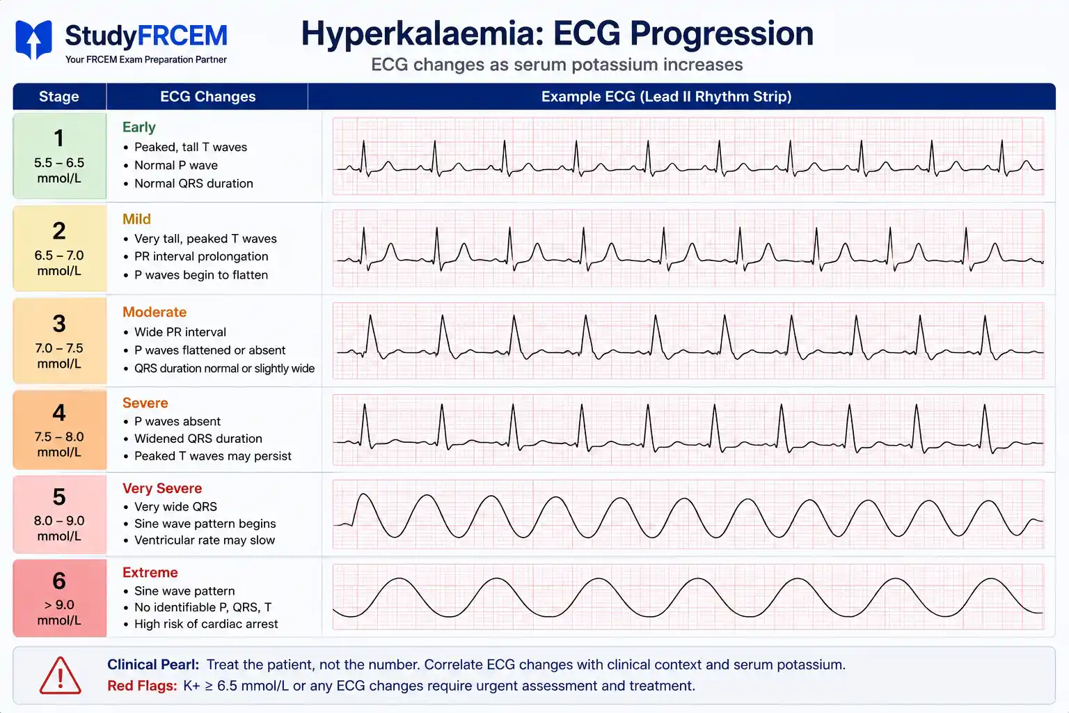
The Progression — In Order
ECG change | Approximate K⁺ level | Clinical implication |
|---|---|---|
Peaked (tented) T waves | 5.5–6.5 mmol/L | Earliest change — easy to miss |
Flattened P waves | 6.0–7.0 mmol/L | Conduction delay emerging |
Prolonged PR interval | 6.5–7.5 mmol/L | AV conduction slowing |
Widened QRS | 7.0–8.0 mmol/L | Ventricular conduction delay — critical threshold |
Sine wave pattern | >8.0 mmol/L | Pre-arrest pattern |
VF / asystole | Variable | Cardiac arrest |
The critical threshold for treatment escalation is QRS widening. Any QRS >100 ms in the context of known or suspected hyperkalaemia is a signal that cardiac arrest may be imminent.
The Key Exam Point: ECG Changes Override the Number
If the K⁺ is 5.8 mmol/L but the QRS is widened — that patient is treated as severe hyperkalaemia emergency, not moderate. The ECG determines urgency, not the laboratory value alone.
Management (Updated UK Kidney Association Guidance)
Cardiac membrane stabilisation: IV calcium gluconate — updated recommended dose is 30 ml of 10% calcium gluconate over 10 minutes (not 10 ml as older resources teach). Use calcium chloride in cardiac arrest or peri-arrest.
Shift K⁺ intracellularly: 10 units soluble insulin in 25g glucose IV infusion; nebulised salbutamol 10–20mg as adjunct
Monitor glucose: at 0, 15, 30 minutes, then hourly for 6 hours — delayed hypoglycaemia is a well-recognised complication
Remove K⁺: dialysis for refractory cases
For full AKI and electrolyte management protocols, see the FRCEM AKI and electrolyte emergencies guide.
5. Posterior MI — The STEMI Hiding in Plain Sight
What It Is
Posterior MI is the STEMI equivalent that hides on a standard 12-lead ECG because posterior changes appear as their mirror image in the anterior leads — not as ST elevation, but as ST depression. Candidates who scan for elevation miss it entirely.
It is caused by occlusion of the right coronary artery or circumflex artery supplying the posterior wall of the left ventricle. It's a true transmural infarction requiring immediate cath lab activation — but it won't trigger an automated STEMI alert on a standard 12-lead, and it won't be caught by a clinician who only looks for elevation.
What to Look For in V1–V3
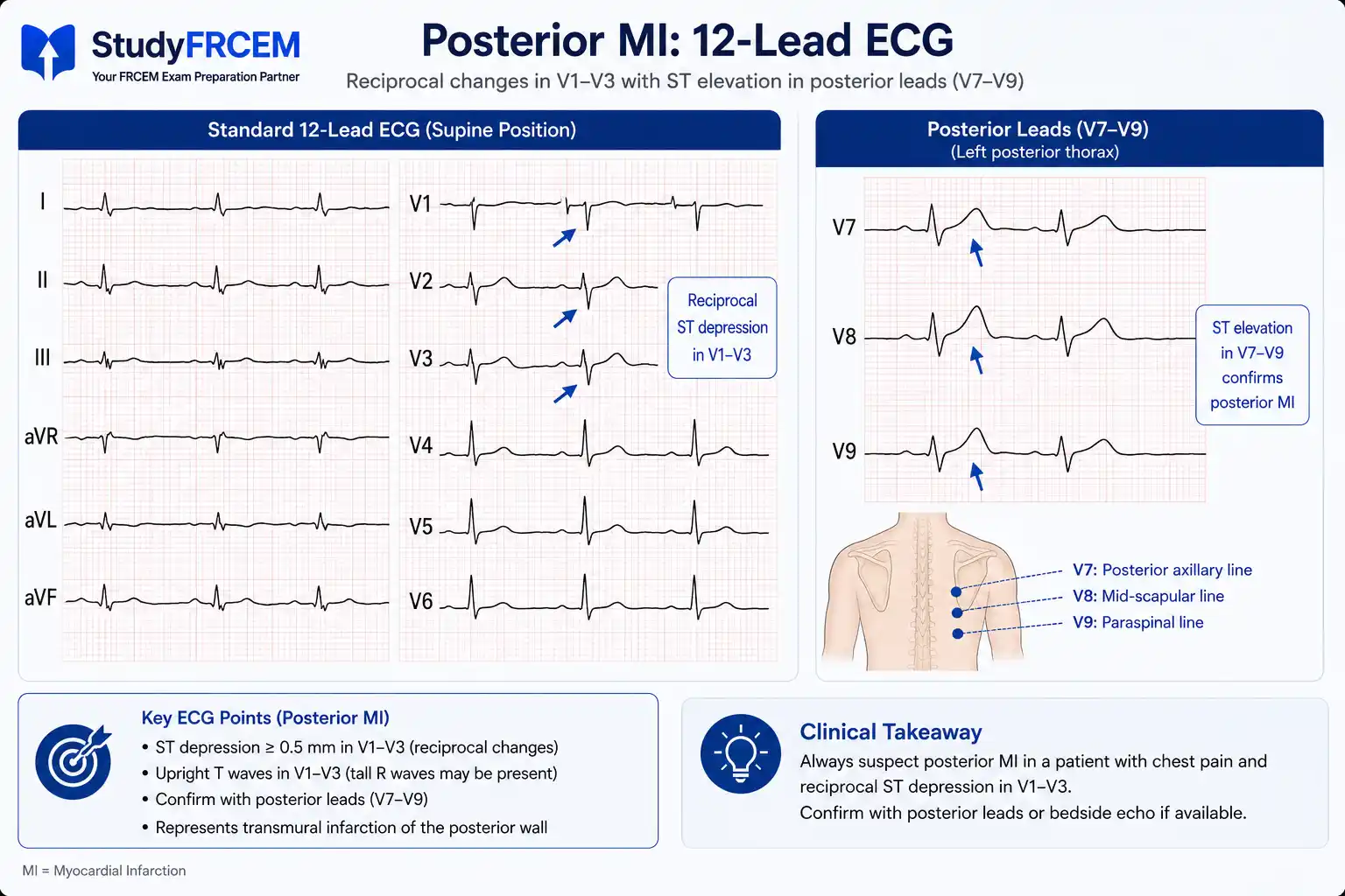
The posterior MI pattern in the anterior leads appears as the reciprocal of posterior ST elevation:
Horizontal ST depression in V1–V3 — not upsloping, not downsloping, but horizontal and significant
Tall, dominant R waves in V1–V2 — R wave taller than S wave (R/S ratio >1 in V2); these are the reciprocal of posterior Q waves
Upright T waves in V1–V2 — normally T waves here are flat or inverted
This combination — horizontal ST depression + tall R waves + upright T waves in V1–V3 — in a patient with chest pain should trigger immediate posterior lead acquisition.
Confirm with Posterior Leads
Apply posterior leads V7, V8, V9 — electrodes placed at the left posterior chest wall. The diagnosis is confirmed by ST elevation ≥0.5 mm in V7–V9. The latest ESC ACS guidelines recommend using right-sided and posterior leads whenever standard leads are inconclusive for inferior or posterior STEMI.
What FRCEM Tests
A scenario with acute chest pain and isolated ST depression in V1–V3 — no obvious ST elevation on the standard 12-lead. The question asks what the diagnosis might be and what investigation should follow.
Wrong answer: Diagnose as NSTEMI and manage conservatively.
Correct answer: Suspect posterior MI. Acquire posterior leads V7–V9. If ST elevation ≥0.5 mm confirmed — immediate cath lab activation as per STEMI protocol. Not NSTEMI management — posterior MI is a STEMI equivalent.
The Five Patterns Side by Side
Pattern | Key ECG finding | Lead location | Urgency | Action |
|---|---|---|---|---|
Wellens' syndrome | Biphasic (Type A) or deep inverted T waves (Type B) | V2–V4 | Urgent (not emergency at presentation) | Cardiology referral, NO stress test |
De Winter's T waves | Upsloping ST depression + tall peaked T waves | V1–V6 (+aVR elevation) | Immediate | STEMI protocol — cath lab now |
Brugada pattern | Coved ST elevation + T wave inversion (Type 1 only) | V1–V3 | Depends on symptoms | Cardiology referral; avoid Na channel blockers; defibrillate VF |
Hyperkalaemia | Peaked T waves → wide QRS → sine wave | Diffuse | Based on ECG changes, not K⁺ level | Calcium gluconate 30ml 10% → insulin/glucose |
Posterior MI | Horizontal ST depression + tall R waves | V1–V3 | Immediate | Posterior leads → STEMI protocol if confirmed |
Common FRCEM Mistakes
1. Discharging a patient with Wellens' pattern because they're pain-free and troponin is near-normal The pain-free, near-normal-troponin presentation is classic Wellens'. Urgent cardiology referral — do not discharge, do not stress test.
2. Ordering a stress test in Wellens' syndrome Explicitly contraindicated — increasing myocardial oxygen demand in a patient with 90%+ proximal LAD stenosis may provoke the very infarction you're trying to prevent.
3. Managing de Winter's pattern as NSTEMI De Winter's is a STEMI equivalent. Upsloping ST depression with tall T waves in precordial leads = proximal LAD occlusion = immediate cath lab.
4. Thinking Brugada Type 2 is diagnostic Only Type 1 (coved pattern) is diagnostic of Brugada syndrome. Type 2 (saddle-back) requires provocation testing. Don't diagnose Brugada syndrome from Type 2 alone.
5. Using ECG potassium level rather than ECG changes to drive treatment urgency ECG changes determine urgency in hyperkalaemia. A wide QRS at K⁺ 6.0 mmol/L is more alarming than peaked T waves at K⁺ 7.0 mmol/L. Treat the ECG, not just the number.
6. Diagnosing posterior MI as NSTEMI based on V1–V3 ST depression alone Check for tall R waves and upright T waves. If the pattern fits, get posterior leads immediately. Horizontal ST depression in V1–V3 is never just "NSTEMI" without ruling out posterior MI first.
Practicing These Pathways With StudyFRCEM
StudyFRCEM's cardiology question bank is built specifically around this kind of exam-style application - covering subtle STEMI equivalents, channelopathies, and metabolic emergencies with ESC- and RCEM-guideline-aligned questions and explanations written by NHS Emergency Medicine consultants. Each question includes a detailed explanation covering not just the correct answer, but exactly why the distractors are wrong, which is the precise reasoning skill the FRCEM SBA is testing.
If you want to see how these high-stakes cardiology scenarios are structured before committing to a plan, the free demo gives you a direct feel for the question style.
Study Strategy
ECG pattern recognition is one of the most transferable skills in FRCEM revision — it appears across resuscitation, cardiology, toxicology, and metabolic emergency questions. Investing time in it pays dividends across multiple question categories.
For these five patterns specifically:
Know Wellens' Type A vs B, and that stress testing is contraindicated
Know de Winter's as a STEMI equivalent — not NSTEMI
Know Brugada Type 1 only is diagnostic; know fever as an ED-specific trigger
Know hyperkalaemia progression in order; know the updated calcium gluconate dose (30ml of 10%)
Know posterior MI's reciprocal changes in V1–V3 and when to get posterior leads
Pair this with the RCEM clinical guidelines cheat sheet to cross-reference management decisions against current guideline recommendations — particularly the updated ESC ACS guidance on P2Y12 inhibitor sequencing and PPCI timing that applies when de Winter's or posterior MI is identified.
These five patterns share a common theme: they look deceptively stable or non-specific, they're easy to dismiss under ED time pressure, and they're precisely the ones FRCEM examiners choose because missing them in real life has catastrophic consequences. Getting fast and accurate at recognising them is one of the highest-return investments you can make in your exam preparation.
For SLO-mapped SBA questions specifically targeting these ECG patterns — with consultant-written explanations for every answer option — register with StudyFRCEM.
Frequently Asked Questions
Are de Winter's T waves actually tested in FRCEM?
Yes, de Winter's is now explicitly listed as a STEMI equivalent in the ESC ACS guidelines, making it a legitimate exam target. It appears in question banks specifically because candidates trained on "ST elevation = STEMI" miss it.
How do I distinguish Wellens' from normal anterior T wave inversions?
Wellens' T wave inversions are deep, symmetrical, and in the context of a patient with recent chest pain or known coronary risk. Normal anterior T wave inversions (in V1–V2) are common and not specifically associated with recent ischaemic symptoms. Context is everything.
Can Brugada pattern be caused by medications in the ED?
Yes, certain drugs can unmask Brugada. The most relevant in the ED: TCA overdose, cocaine, some antipsychotics (especially phenothiazines), and some anaesthetic agents. This overlaps with toxicology questions. A patient who takes TCAs and has right precordial ST changes may have either TCA toxicity or Brugada unmasking - or both.
What if the posterior leads are negative but I still strongly suspect posterior MI?
If clinical suspicion is high (mechanism, risk factors, V1–V3 changes), discuss with a cardiologist before dismissing it as NSTEMI. A negative posterior lead result on a poor-quality trace in a symptomatic high-risk patient warrants further evaluation - repeat ECG and serial troponin minimum.
How many of these patterns appear in a single FRCEM sitting?
There's no fixed rule, but ECG-based questions across the cardiology, resuscitation, and toxicology themes typically account for 8–12 questions per sitting. One or two of these specific patterns will appear - and they're reliable discriminators between candidates who have specifically studied them and those who haven't.