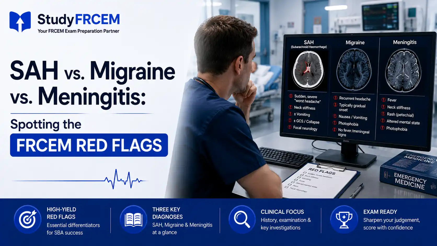

A patient walks into your emergency department with a severe headache. They're in their 40s, otherwise fit, and the headache came on suddenly. It could be a migraine — or it could be a subarachnoid haemorrhage that kills them if you send them home.
This is the clinical scenario the FRCEM SBA loves most. Not because it's rare, but because the differential is genuinely hard, the consequences of getting it wrong are catastrophic, and the guidelines give you very specific tools to navigate it. Candidates who know those tools — the exact Ottawa SAH criteria, the NICE NG240 meningitis red flags, the distinction between sentinel headache and migraine — score reliably on these questions. Those who rely on vague clinical intuition get the wrong answer.
This guide covers exactly what FRCEM tests when it pits SAH, bacterial meningitis, and migraine against each other: the red flags, the investigation pathway, and the management decisions that distinguish each condition.
Why This Differential Matters So Much in the SBA
Headache questions account for a predictable number of marks across SLO1 and SLO4. They're designed to test whether you:
Know which features demand investigation regardless of how "typical" the presentation looks
Understand the correct investigation sequence — and crucially, when a negative CT does not clear the patient
Apply the right management decisions at the right speed
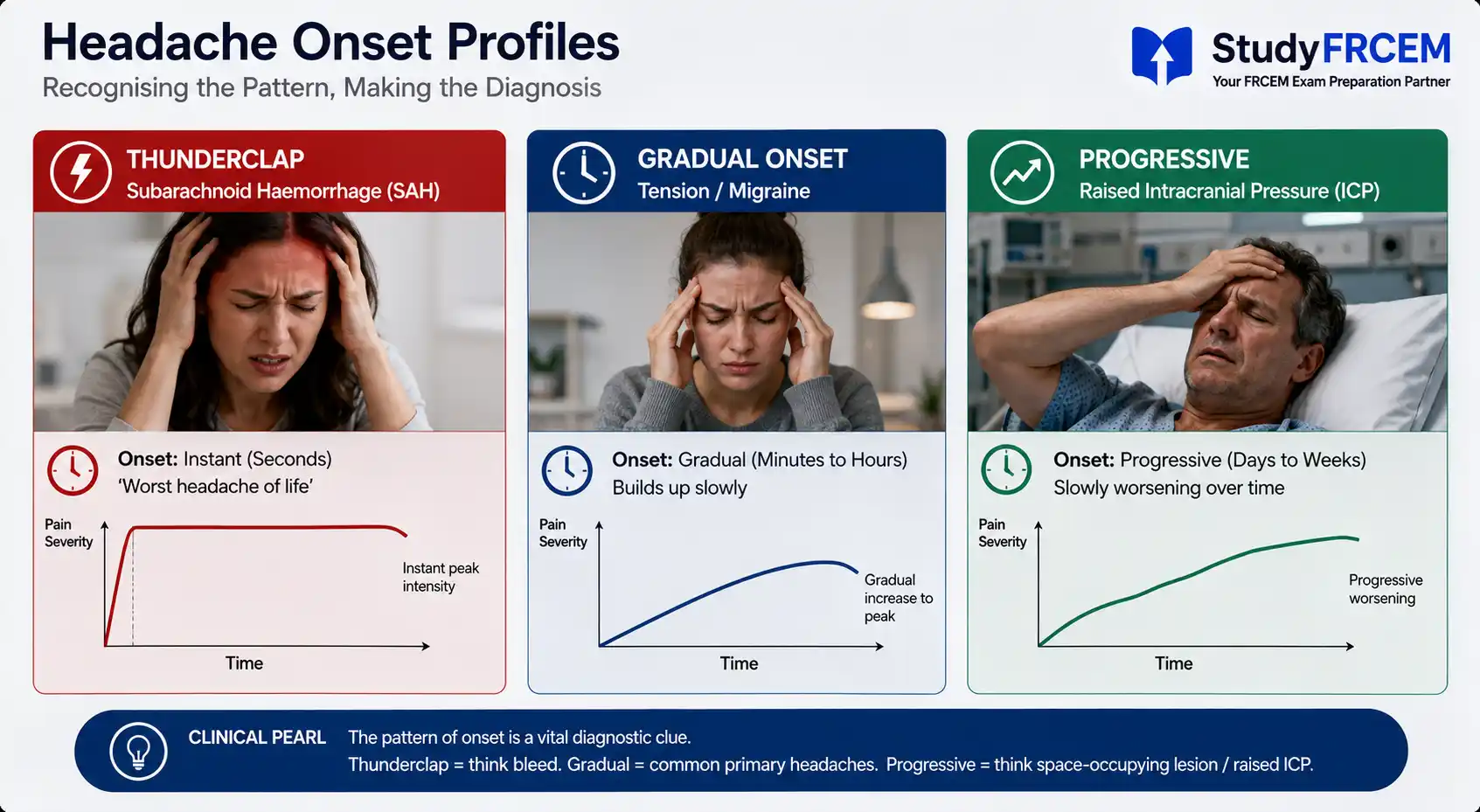
The classic trap is the patient with a headache that "sounds like their usual migraines" — except this one was instantaneous, while vomiting, at maximal severity from the first second. That's a thunderclap headache. That's SAH until proven otherwise.
Subarachnoid Haemorrhage: The Diagnosis You Cannot Miss
The Thunderclap Headache
SAH caused by ruptured cerebral aneurysm presents with a thunderclap headache — defined as a severe headache that reaches maximal intensity instantly (or within seconds to minutes) at onset. Patients often describe it as "the worst headache of my life" — but that phrase alone isn't enough. The speed of onset is what matters.
In the majority of cases of missed spontaneous SAH, the most common mistake was not considering the diagnosis and not ordering a non-contrast CT head. Spontaneous SAH has a spectrum of disease presentation, and clinicians cannot rely on the "classic presentation" as the only trigger for ordering imaging.
This is the exam point: SAH doesn't always look textbook. Some patients are alert and seemingly stable. Some describe a headache that has partially improved. The investigation threshold is clinical, not determined by how unwell the patient appears.
Red Flags for SAH — Know Every One
The following features, in the context of acute severe headache, should prompt immediate SAH workup:
Thunderclap onset — maximum intensity at or within seconds of onset
Exertional onset — headache starting during physical activity, sexual activity, or straining
Occipital location — particularly if radiating to the neck
Associated loss of consciousness — even brief or witnessed syncope
Neck stiffness or meningism — though this may take hours to develop
New neurological deficit — focal signs suggest complications (hydrocephalus, vasospasm)
Sentinel headache — a severe headache in the days or weeks prior ("warning leak"), which patients may dismiss
The sentinel headache is particularly high-yield for FRCEM. A patient who comes in with a moderately severe headache that is "different from usual" and describes a similar headache two weeks ago that "went away on its own" — that history should make you concerned about a warning leak preceding a full SAH. Do not be reassured by the fact they look well now.
The Ottawa SAH Rule
The Ottawa SAH Rule is the validated clinical decision tool for identifying patients with non-traumatic headache who require CT. The Ottawa SAH rule is a clinical decision tool to aid in the decision for computed tomography of the head in patients attending an emergency department with acute non-traumatic headache.
Investigate for SAH (CT head required) if any one of the following is present:
Age ≥40
Neck pain or stiffness
Witnessed loss of consciousness
Onset during exertion
Thunderclap onset (instant maximal severity)
Limited neck flexion on examination
The Ottawa Rule is designed to be highly sensitive — it casts a wide net deliberately, because missing SAH is the catastrophic outcome. It is not designed to be specific. A positive Ottawa Rule means "investigate," not "this patient has SAH."
FRCEM application: If a question presents an acute severe headache with any Ottawa criterion and asks what investigation is required, the answer is immediate non-contrast CT head.
CT Head: The 6-Hour Rule
CT head sensitivity for excluding SAH at under 6 hours from symptom onset is approximately 0.995 (95% CI 0.941–1.000). At more than 6 hours, pooled sensitivity falls to 0.94.
This is one of the most clinically important and most tested numbers in SAH diagnosis:
CT within 6 hours of onset + negative result = SAH effectively excluded in most patients
CT more than 6 hours after onset + negative result = does NOT exclude SAH
If CT is negative but the patient presented more than 6 hours after headache onset, or if clinical suspicion remains high despite a negative early CT, lumbar puncture is required.
LP continues to be an important diagnostic test that will confirm a diagnosis of aneurysmal SAH in a small but significant number of patients with thunderclap headache. The NICE guideline states that a sudden severe headache typically peaking in intensity within 1 to 5 minutes is a red-flag symptom of SAH, and that if a CT head scan is non-diagnostic, LP should be performed.
Lumbar Puncture for SAH
LP must be performed at least 12 hours after headache onset — this allows time for xanthochromia (yellow discolouration from haemoglobin breakdown products) to develop in the CSF.
What to look for:
Xanthochromia — the most specific finding; detected visually or by spectrophotometry
Uniformly bloodstained CSF across all three tubes (distinguishes from traumatic tap, where blood clears in successive tubes)
Elevated opening pressure
FRCEM trap: If LP is performed too early (before 12 hours), xanthochromia may not yet have developed and a negative result is unreliable. The correct answer to "when should LP be performed to investigate SAH?" is at least 12 hours after onset.
SAH Management
Once SAH is confirmed or strongly suspected:
Urgent neurosurgical referral
IV access, cardiac monitoring (SAH causes ECG changes — peaked T waves, QT prolongation, deep T wave inversions — that mimic ACS)
Analgesia and antiemetics
Nimodipine 60mg orally every 4 hours — reduces cerebral vasospasm and improves neurological outcome; initiated as soon as SAH is confirmed
BP control — avoid hypotension (risks ischaemia) and hypertension (risks re-bleeding); target SBP <160 mmHg typically, guided by neurosurgical advice
Avoid LP if CT shows large haematoma or obstructive hydrocephalus
FRCEM testing: Questions on SAH management often test nimodipine (vasospasm prevention), timing of LP (≥12 hours), and the ECG changes that can mislead into an ACS diagnosis.
Bacterial Meningitis: The Diagnosis That Cannot Wait
NICE NG240 — Updated Guidance
NICE has published a new guideline (NG240) on the diagnosis and management of bacterial meningitis and meningococcal disease. The guidance updates and replaces the previous guideline CG102, published in June 2010.
This is a recently updated guideline and directly relevant to FRCEM — know what's changed.
Red Flag Combination for Bacterial Meningitis
Bacterial meningitis should be strongly suspected in adults, children and babies with the "red flag combination" — fever, headache, neck stiffness, and altered level of consciousness or cognition (including confusion or delirium).
Not all four features need to be present. The key principle is: any combination of fever + headache + neck stiffness warrants urgent consideration of bacterial meningitis, particularly with any alteration in conscious level.
Classical triad: Fever + neck stiffness + photophobia/headache
Additional features:
Photophobia
Non-blanching petechial or purpuric rash — suggests meningococcal septicaemia; a medical emergency
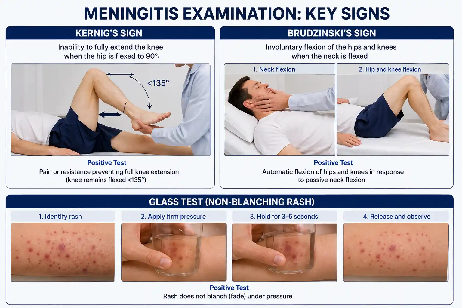
Kernig's sign — inability to extend the knee with hip flexed 90°
Brudzinski's sign — involuntary knee flexion when neck is flexed
Seizures
Bulging fontanelle in infants
High-pitched cry in infants
The rash: A non-blanching rash in the context of fever and systemic illness is meningococcal disease until proven otherwise. Do the glass test — press a glass against the skin; a petechial rash that doesn't blanch under pressure indicates haemorrhage into the skin from meningococcaemia. This is a clinical emergency — do not wait for investigations before treating.
Investigation: When to Do LP and When Not To
If it is safe to do so and will not cause a clinically significant delay to starting antibiotics, perform a lumbar puncture before giving antibiotics.
However, LP is contraindicated in the following circumstances:
Signs of raised intracranial pressure (papilloedema, reduced GCS, focal neurological deficits, Cushing's triad)
Coagulopathy or anticoagulation
Septic shock
Infection at the LP site
Recent seizure
If LP is contraindicated: start antibiotics immediately, then obtain CT head before LP.
Blood cultures must be taken before antibiotics — if this will cause any delay in giving antibiotics, take the cultures as rapidly as possible but do not delay treatment for them.
Antibiotic Treatment — NICE NG240
Give ceftriaxone as the first-line hospital treatment while the causative organism is unknown; add amoxicillin if there are risk factors for Listeria monocytogenes.
Risk factors for Listeria (requiring amoxicillin addition): age >60, immunocompromised, pregnancy, alcoholism.
Course length:
Meningococcal meningitis: 5 days (shortened course per new NG240 evidence)
Pneumococcal meningitis: 10–14 days
Listeria: 21 days
Penicillin allergy: chloramphenicol (discuss with infection specialist).
Dexamethasone — Important Rule
For people over 3 months with strongly suspected or confirmed bacterial meningitis, give intravenous dexamethasone.
Dexamethasone should be continued if the cause is found to be pneumococcus or Haemophilus influenzae type b, but stopped if the causative agent is any other organism. However, corticosteroids should not routinely be given to people with meningococcal disease (except in meningococcal septic shock not responding to high-dose vasoactive agents).
Timing: Give dexamethasone with or just before the first dose of antibiotics. If dexamethasone is delayed for more than 12 hours after the start of antibiotics, get advice from an infection specialist and decide whether dexamethasone should still be given.
FRCEM key points on dexamethasone:
Give it in bacterial meningitis (age >3 months) — specifically before or with antibiotics
Continue only if pneumococcal or H. influenzae b cause confirmed
Do not give routinely in meningococcal disease — this is explicitly stated in NG240 and is a common trap question
Migraine: Recognising What It Is — and What It Isn't
Migraine is common, and FRCEM tests your ability to diagnose it confidently while maintaining the right level of suspicion for its dangerous mimics. The key is recognising both the positive features of migraine and the red flags that should override that diagnosis.
Positive Migraine Features
Gradual onset — typically building over 30 minutes to hours (never instantaneous)
Unilateral throbbing/pulsating headache
Moderate to severe intensity, worsening with activity
Nausea and/or vomiting
Photo- and phonophobia
Preceding aura (in approximately 25% of cases) — visual disturbances (fortification spectra, scotoma), sensory changes, speech disturbance, lasting 20–60 minutes
Previous identical episodes — a history of recurrent identical attacks is the most reassuring feature
The Migraine Red Flags That Override the Diagnosis
Even in a patient with a known migraine history, these features demand investigation:
Red flag | Concern |
|---|---|
Thunderclap onset | SAH |
First or worst headache ever | SAH |
Headache "different from usual" | SAH, space-occupying lesion |
Fever + neck stiffness | Meningitis |
New focal neurological deficit | Stroke, SOL, complicated migraine |
Onset after 50 | Temporal arteritis, space-occupying lesion |
Progressive worsening over weeks | Space-occupying lesion |
Onset during exertion or sex | SAH |
Associated jaw claudication, scalp tenderness | Temporal arteritis (ESR, CRP, temporal artery biopsy) |
FRCEM principle: Never diagnose migraine for the first time without excluding SAH if the onset was thunderclap, the headache is the worst ever, or there are any of the above red flags. A diagnosis of migraine in the ED is a diagnosis of exclusion.
Migraine Management in the ED
For established migraine without red flags:
Aspirin 900 mg or ibuprofen 400–600 mg — first-line analgesics
Metoclopramide 10 mg IV/IM — antiemetic and enhances analgesic absorption; also has mild antimigraine properties
Triptans (e.g. sumatriptan 50–100 mg oral, or 6 mg SC) — specific for migraine with or without aura; first choice if analgesics fail or severe
Avoid opioids — not recommended for migraine; cause medication overuse headache and don't address the underlying pathophysiology
Status migrainosus (migraine lasting >72 hours): consider IV prochlorperazine, IV valproate, or a short course of oral dexamethasone — discuss with neurology.
The Three-Way Differential: Key Distinguishing Features
Feature | SAH | Bacterial Meningitis | Migraine |
|---|---|---|---|
Onset | Thunderclap — instantaneous | Subacute (hours–days) | Gradual (minutes–hours) |
Severity | Severe, immediate peak | Progressive, severe | Moderate–severe, builds up |
Fever | Absent initially | Yes | No |
Neck stiffness | Develops over hours | Early sign | No |
Rash | No | Petechiae/purpura (meningococcal) | No |
Photophobia | Yes | Yes | Yes |
Previous identical episodes | No (first episode) | No | Yes (recurrent) |
LOC | May occur | Late feature | Rarely |
Sentinel headache | Yes (warning leaks) | No | No |
Key investigation | CT + LP if >6h or high suspicion | Blood cultures + LP (if safe) | Clinical diagnosis |
Key treatment | Nimodipine + neurosurgery | IV ceftriaxone + dexamethasone | NSAIDs + antiemetic + triptan |
Common FRCEM Mistakes
1. Dismissing SAH because the patient looks well SAH can present in alert, neurologically intact patients. Clinical appearance does not determine the investigation threshold. Ottawa criteria do.
2. Not performing LP because CT was negative If CT is negative and was performed more than 6 hours after onset — or clinical suspicion remains high — LP is mandatory. A negative CT alone does not rule out SAH beyond the 6-hour window.
3. Performing LP too early for SAH LP for SAH must be at least 12 hours after onset to allow xanthochromia to develop. Earlier LP can produce a false-negative result.
4. Giving dexamethasone in meningococcal disease routinely NG240 explicitly states do not routinely give corticosteroids in meningococcal disease. Dexamethasone is for bacterial meningitis — and should be stopped if the organism is not pneumococcus or H. influenzae b.
5. Diagnosing migraine on first presentation with thunderclap onset First-ever thunderclap headache is SAH until proven otherwise. Never accept a migraine diagnosis at face value when the onset feature is thunderclap.
6. Delaying antibiotics to obtain LP in meningitis If LP is contraindicated — or will cause a clinically significant delay — start IV ceftriaxone immediately. Blood cultures first if rapid, but antibiotics must not be delayed by more than a few minutes.
Practicing These Pathways With StudyFRCEM
StudyFRCEM's neurology question bank is built specifically around this kind of exam-style application—covering SAH, bacterial meningitis, and complex migraines with NICE- and RCEM-guideline-aligned questions and explanations written by NHS Emergency Medicine consultants. Each question includes a detailed explanation covering not just the correct answer, but why the distractors are wrong, which is exactly the reasoning skill the FRCEM SBA is testing.
If you want to see how these scenarios are structured before committing to a plan, the free demo gives you a direct feel for the question style.
Study Strategy
Headache differentials appear in multiple SLO domains. The specific clinical reasoning tested — investigation sequencing, Ottawa criteria, meningitis management steps — rewards focused revision over broad reading.
Priority areas:
Ottawa SAH Rule criteria (6 specific features)
CT 6-hour rule and xanthochromia LP timing (12 hours minimum)
Nimodipine in SAH management
NICE NG240 meningitis: red flag combination, ceftriaxone, dexamethasone use and when to stop it
Non-blanching rash as a clinical emergency
Migraine red flags that demand investigation
This topic dovetails directly with the FRCEM stroke and TIA SBA guide — both cover neurological emergencies where time-critical investigation decisions determine outcome, and the clinical reasoning transfers between them. For the broader guideline framework that contextualises these decisions, the RCEM clinical guidelines cheat sheet cross-references NICE NG240 alongside the other major updated guidelines.
The headache differential is one of the highest-stakes clinical decisions in emergency medicine — and one of the most reliably tested in the FRCEM SBA. Master the Ottawa SAH criteria, the 6-hour CT rule with its LP consequences, and the NICE NG240 meningitis management sequence, and these questions move from anxiety-inducing to genuinely scorable.
For SLO-mapped SBA questions covering SAH, bacterial meningitis, and migraine with detailed guideline-based explanations, register with StudyFRCEM.
Frequently Asked Questions
Does the Ottawa SAH Rule apply to all headaches?
No, it applies specifically to patients presenting to the ED with acute non-traumatic headache, who are neurologically intact. It is not validated in patients with GCS <15, known intracranial pathology, or papilloedema.
Is xanthochromia always visual?
Visual xanthochromia (yellow tinge to CSF visible to the naked eye) is the bedside assessment. Spectrophotometry is more sensitive and is the recommended laboratory method in the UK - it measures oxyhaemoglobin and bilirubin separately. Some centres use spectrophotometry routinely; others rely on visual inspection. For FRCEM purposes, xanthochromia is the key finding regardless of detection method.
What ECG changes does SAH cause?
SAH can cause peaked T waves, deep symmetric T wave inversions, QTc prolongation, and ST changes that mimic ACS. Always get a 12-lead ECG in confirmed or suspected SAH - and don't let a "normal" ECG reassure you, as changes can be delayed.
Should I give pre-hospital antibiotics for suspected meningitis?
NG240 notes that the priority is transfer to hospital. If antibiotics are given outside hospital, ceftriaxone is preferred (more active, though less commonly available pre-hospital); benzylpenicillin is recommended as it is more commonly available in pre-hospital settings.
How many headache/neurological questions appear in FRCEM?
Approximately 6–10 across SLO1 and SLO4 per sitting, covering SAH, meningitis, migraine, stroke, and space-occupying lesions. The SAH investigation pathway and meningitis management steps appear in most sittings.